The University of British Columbia
UBC - A Place of Mind
The University of British Columbia
Faculty of MedicineMoore Lab | Department of Cellular & Physiological Sciences
  • Home
  • Methods
  • Publications
  • Contact
  • Our Team
  • Model of a Rat Ventricular Myocyte Dyad.

    Thermo Scientific Amira Software were used to prepare this model from the 3D reconstructed tomogram of the dyad. Read More

  • Localization, Distribution and NND

    Localization, distribution and center-to-center nearest neighbour distance (NND) measurements of RyR2 tetramers on the surface of the jSR in a mouse ventricular myocyte using Electron Tomography (3D Electron Microscopy). Read More

  • SR content by 20mM caffein application

    SR Calcium Content

    Measuring Sarcoplasmic Reticulum Calcium content by appliny 20 mM Caffein to permibilized ventricular mycytes. Read More

  • Superresolution Image of the Distribution of RyR2

    Superresolution images of the surface of isolated cardiomyocytes from notmal rat labeled for the ryanodine receptor Read More

  • Transmission electron micrograph of two surface junctions in rat ventricle

    Read More

Regulating Cardiac Contractile Force

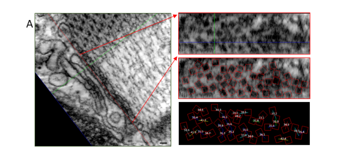
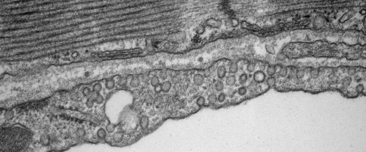
The type-2 cardiac ryanodine receptor (RyR2) is a Ca2+-activated Ca2+ ion channel located in the junctional sarcoplasmic reticulum (jSR) whose primary function is to regulate the amount, and the rate, of Ca2+ released from the jSR. Ca2+ release is the primary determinant of cardiac contractile force. Each jSR is decorated with between one and several hundred RyR2 that affect each other’s open probability through Ca2+-induced Ca2+ release (CICR) and through allosteric interactions. Recent and exciting data from the lab has shown that RyR2 channels are mobile and can interact with their neighbours in multiple ways. The interactions are dynamic and dependent on post-translational modifications and ligands. This opens an unprecedented higher-level type of regulation, whereby the activity of RyR2, and hence the amount of Ca2+ released, could be dictated by making and breaking allosteric coupling between RyR2 channels.

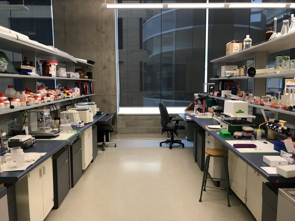
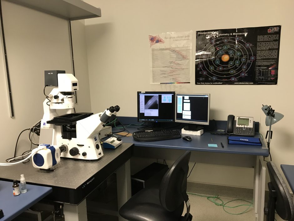
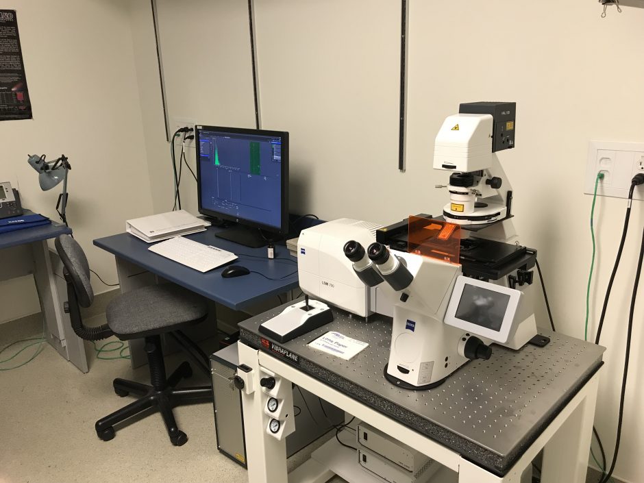
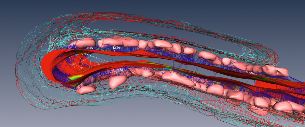
We are investigating RyR2 position and function in both normal and diseased tissue using a variety of cutting-edge and established techniques, including transmission electron microscopy, electron tomography, confocal and deconvolution microscopy, 3D superresolution immunofluorescence microscopy, transgenic mice, Ca2+ spark and Ca2+ transient analyses as well as biochemical approaches.


Correlative Microscopy; Electron Tomography and Calcium Sparks Imaging
direct Stochastic Optical Reconstruction Microscopy (dSTORM)

Moore Laboratory

Moore Lab | Department of Cellular & Physiological Sciences
Faculty of Medicine
2350 Health Sciences Mall
Vancouver, BC Canada V6T 1Z3
Tel 604 822 7719
Back to top
The University of British Columbia
  • Emergency Procedures |
  • Terms of Use |
  • Copyright |
  • Accessibility